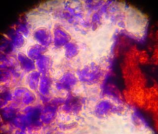
After looking at the saffron stained slide for a while we decided to try adding aniline blue. Unfortunately the cells were unable to withstand the separation of cover slip and slide, and large chunks came free of the cover slip. Here you can see some of the cells stained blue, and some of the connective material stained red. Things were such a mess that it was difficult to see any patterns in these slides. We have begun formulating plans for more gentle staining in the future however.


3 Comments:
I love these pictures. They look great. Hope it has some meaning however!!
I love these pictures. They look great. Hope it has some meaning, however!!
It seems that we have good evidence that large collagen fibers are not yet present in these cultures. The amorphous material that we see in the photos above and below is probably proteoglycan -- aggrecan. We may be able to see collagens just beginning to be visible as short fibers, if we stain with anti-collagen II.
SYNTHESIS AND EXTRACELLULAR DEPOSITION OF FIBRONECTIN IN CHONDROCYTE CULTURES. 1978. WALTRAUD DESSAU, JOACHIM SASSE, RUPERT TIMPL, FRANZ JILEK, and KLAUS VON DER MARK J. CELL BIOLOGY 342-355.
Post a Comment
<< Home