Monday, February 14, 2005
Previous Posts
- This is the same location as the previous picture,...
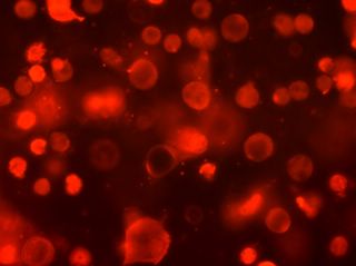
- I tried some SYPRO Ruby protein stain on our cells...
- This is safranin and bradford. The bradford picke...
- Here I tried to combine safranin and aniline blue....
- This is safranin at 40x. It seems to work pretty ...
- This is a Haematoxylin and aniline blue. I found ...
- Another congo red at 40x. This shot is exactly th...
- This is congo red at 40x. The congo red didn't se...
- Another alcian blue at 40x. I thought this one lo...
- More alcian blue at 20x.
Lab Links
ACofI Cartilage Page
Dr. Ayers
Tshering
Alan
Becca
Chadwick
Inez
Sara


1 Comments:
I think that if you look in the lower right hand quadrant you can see the same staining pattern as found with colII antibody. The ruby seems to be staining the collagen and the perlecan.
Post a Comment
<< Home