Saturday, March 05, 2005
Previous Posts
- Here is a spot at 10x
- Last. I would suggest opening all of these shots ...
- next
- next
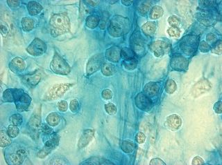
- These are cells grown in 99% Optimem, and 1% ITS. ...
- Another thick fibril.
- 40x with different focus. The big hook-shapped fi...
- And again at 40x.
- Same spot at 20x
- The ITS cells were not nearly as thick across the ...
Lab Links
ACofI Cartilage Page
Dr. Ayers
Tshering
Alan
Becca
Chadwick
Inez
Sara


1 Comments:
It seems to me that these cells have clear areas around them similar to the perlecan stained regions found in comparing the fluorescent and phase contrast images in the Gallery. Does the Alcian Blue stain chondroitin sulfate, but not heparan sulfate?
Post a Comment
<< Home