Monday, February 14, 2005
Previous Posts
- Another at 40x. It was really hard to get anythin...
- Here are some photos at 40x.
- This is what it looked like at 10x. These cells w...
- This is the same location as the previous picture,...
- I tried some SYPRO Ruby protein stain on our cells...
- This is safranin and bradford. The bradford picke...
- Here I tried to combine safranin and aniline blue....
- This is safranin at 40x. It seems to work pretty ...
- This is a Haematoxylin and aniline blue. I found ...
- Another congo red at 40x. This shot is exactly th...
Lab Links
ACofI Cartilage Page
Dr. Ayers
Tshering
Alan
Becca
Chadwick
Inez
Sara


1 Comments:
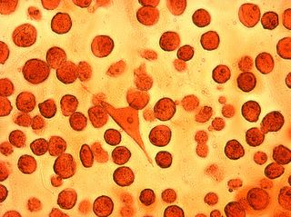
I think that the safranin shows some fiber-like structures that are obscured by the thickness of the matrix. Why does the Ruby not stain? Is it the peculiar amino acid composition of the collagens? We will know when we switch to antibody staining.
Post a Comment
<< Home