ECM Research

Monday, February 14, 2005


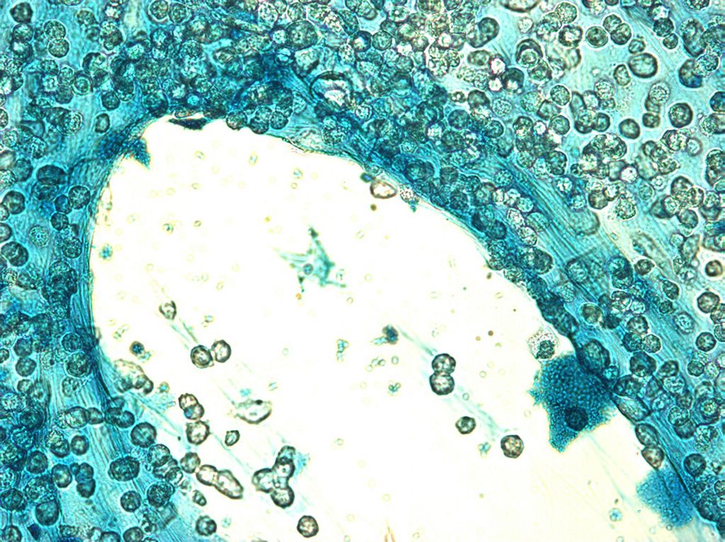
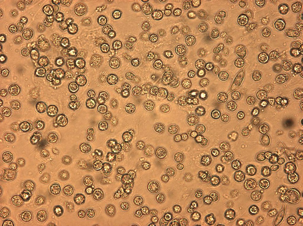
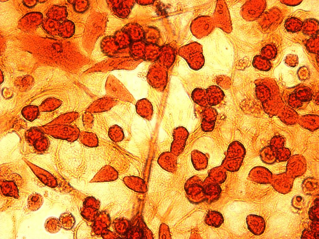
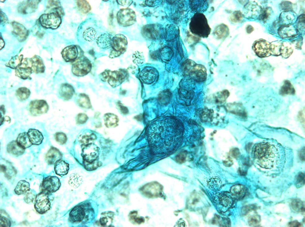
Here is a comparison shot taken with safranin. I had to look long and hard to find this non-round cell. Posted by Hello


Another at 40x. It was really hard to get anything to focus very well when in flourescent mode. Posted by Hello


Here are some photos at 40x. Posted by Hello


This is what it looked like at 10x. These cells were from 1-27-2005 and are very old. They are very thick, and it seemed like a larger percentage than normal were round instead of odd shapped. Posted by Hello


This is the same location as the previous picture, but with phase constrast. Both are are taken with the 20x lense. Posted by Hello


I tried some SYPRO Ruby protein stain on our cells. If the pictures are any indication, we have no protein in the ECM. On the plus side, I learned how to use the fluorescent scope. It is impressive how quickly the stain gets bleached out! Posted by Hello

Tuesday, February 08, 2005


This is safranin and bradford. The bradford picked up some things, but not many. it also seemed to somewhat remove the safranin to a certain extent. Posted by Hello


Here I tried to combine safranin and aniline blue. The results were not very spectacular. This is at 20x. Posted by Hello


This is safranin at 40x. It seems to work pretty consistently so I did one coverslip with it sort of as a control. The results look more or less like those from previous safranin stained samples. The optics are a bit different however, as is the exposure on the camera attached to the microscope. Posted by Hello


This is a Haematoxylin and aniline blue. I found a wonderful example of this being used in which it was claimed that elastic fibers stained dark purple, and collagen blue. It turns out that haematoxylin staining is much more complicated that I initially hoped however. According to >this site, haematoxylin is “not a dye…[and has] little or no affinity for tissue elements and requires an inorganic ion to act as a ‘go between’ between the dye and the tissue.” Well, NO WONDER it didn’t work! Apparently a ferric salt can be used in some way (further research is needed here).
 Posted by Hello


Another congo red at 40x. This shot is exactly the same as the last one, except the objective has been moved back a bit. This shows the multi-layered nature of these cells. For the most part, the ones which are against the glass seem to be dividing and strangly shaped while those which are suspended in the ecm are much rounder. Posted by Hello


This is congo red at 40x. The congo red didn't seem to stain the ecm well, but did a good job of making the cells show up. When the objective is moved towards the slide these cells come into focus. Posted by Hello


Another alcian blue at 40x. I thought this one looked especially interesting. Posted by Hello


More alcian blue at 20x. Posted by Hello


This is alcian blue stain at 40x. Apparently it is traditionally used to stain mucopolysaccharides or glycosaminoglycans, and is a cationic dye which is attracted to anionic sites on polysaccharides. Posted by Hello

More Staining (now with the nicer scope)

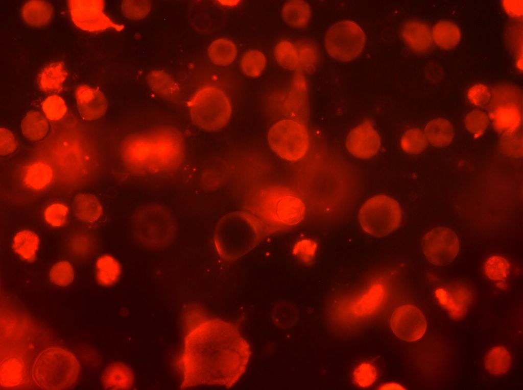
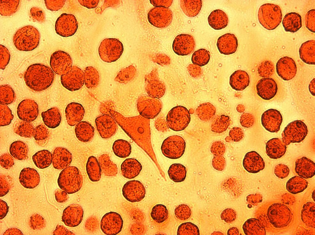
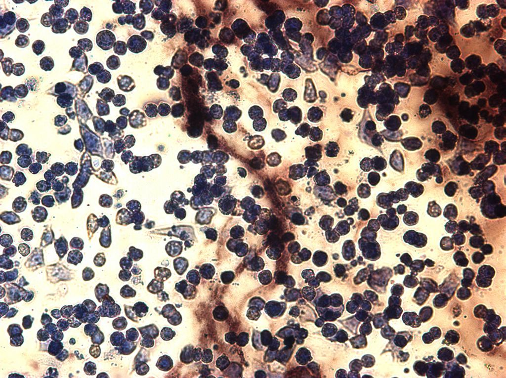
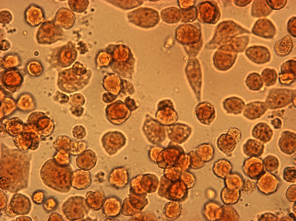
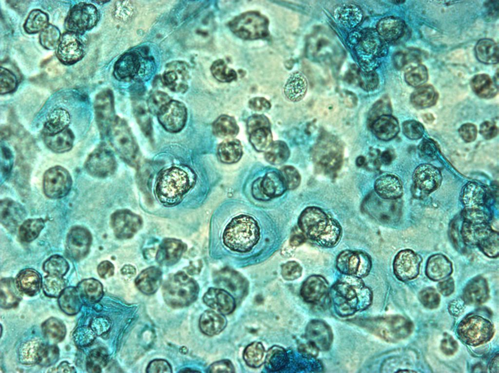
Using the same fixing/staining procedure as in the last set of stains, six new staining experiments were carried out. This time the photos were taken using a camera mounted on a somewhat higher end microscope. All staining and photographing was done on 2-03-2005. Sample pictures are above.